Ct Anatomy Pelvis Muscles - Female pelvis anatomy | free axial cross sectional anatomy ... : Included within the chart are gorgeous illustrations of the pelvic diaphragm, sphincter muscles, gluteus maximus.
Dapatkan link
Facebook
X
Pinterest
Email
Aplikasi Lainnya
Ct Anatomy Pelvis Muscles - Female pelvis anatomy | free axial cross sectional anatomy ... : Included within the chart are gorgeous illustrations of the pelvic diaphragm, sphincter muscles, gluteus maximus.. The video covers the most. This mri male pelvis axial cross sectional anatomy tool is absolutely free to use. Figure 6.4 • ct scan of pelvis: How to view anatomical labels. Architectural differences in the bony pelvis of women with and without pelvic floor disorders.
Techniques should provide image quality consistent with the diagnostic needs. Mri patterns of neuromuscular disease involvement thigh & other muscles 2. These slides were taken from wikiradiography (wetpaint) here. The muscles of the pelvis, hip and buttock anatomical chart shows how each muscle in this area of the body works with the others, and the various minor systems within the major ones. It is a powerful hip extensor that acts to bring the thigh in a straight line with the pelvis.
Abdominal CT anatomy | Radiology Key from radiologykey.com Architectural differences in the bony pelvis of women with and without pelvic floor disorders. In the back the posterior superior iliac spines are surrounded by muscles and flank fat. Intravenous contrast has been given. How to view anatomical labels. Innervation of the female levator ani muscles. Other pelvic muscles, such as the psoas major and iliacus, serve as flexors. Related online courses on physioplus. Free and interactive atlas of the human anatomy.
Use the mouse scroll wheel to move the images up and down alternatively use the tiny arrows (>>) on both side of the image to move the images.
Involved early gray = muscle: Innervation of the female levator ani muscles. If you want to learn how to read ct scans of the abdomen and pelvis proficiently, this video is an excellent starting point. This tool provides access to a ct atlas in the axial plane, allowing the user. The anterior part is called the pelvic girdle which is composed of. Ct of the abdomen axial anatomy. Figure 6.4 • ct scan of pelvis: Related online courses on physioplus. Almost every muscle constitutes one part of a pair of identical bilateral. Furthermore, the pelvis protects the pelvic and abdominopelvic viscera. Included within the chart are gorgeous illustrations of the pelvic diaphragm, sphincter muscles, gluteus maximus. Free and interactive atlas of the human anatomy. Some of the most important include the major digestive organs, the intestines.
Functional anatomy of the male. The muscles of the pelvis, hip and buttock anatomical chart shows how each muscle in this area of the body works with the others, and the various minor systems within the major ones. This video details comprehensive anatomy of the abdomen with coronal correlation.please visit us at www.pulseradiology.com. Ct of the abdomen axial anatomy. Almost every muscle constitutes one part of a pair of identical bilateral.
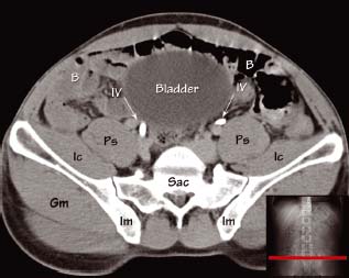
CT Abdomen and Pelvis Coronal Anatomy in the Male ... from i.pinimg.com Free and interactive atlas of the human anatomy. Protocols may be prepared by clinical indication and anatomy to be imaged. Intravenous contrast has been given. Axial section through male bladder. 3 enumerate the muscles of true pelvis. 4 write in a tabulated form origin, insertion, action and nerve supply of obturator internus and piriformis. Functional anatomy of the male. Furthermore, the pelvis protects the pelvic and abdominopelvic viscera.
A variably thick muscular membrane called a diaphragm coccygeus and levator the muscles are attached along the inner walls of the true pelvis to a condensed area of the master the pelvic floor muscles anatomy with our video tutorials, quizzes, labeled diagrams, and articles
This tool provides access to a ct atlas in the axial plane, allowing the user. Anatomy of the muscular system. Microscopic anatomy of skeletal muscle. Protocols may be prepared by clinical indication and anatomy to be imaged. This is a table of skeletal muscles of the human anatomy. Axial section through male bladder. The muscles of the pelvis, hip and buttock anatomical chart shows how each muscle in this area of the body works with the others, and the various minor systems within the major ones. Anatomy of pelvis explained and the most important part pelvic floor consisting of pubococcygeus, puborectalis, and iliococcygeus. Normal ct abdomen/pelvis (without labels). These muscles, including the gluteus maximus and the hamstrings, extend the thigh at the hip in support of the body's weight and propulsion. It is a powerful hip extensor that acts to bring the thigh in a straight line with the pelvis. Anatomy of the abdominal cavity and the male pelvis: Functional anatomy of the male.
Ct abdomen ct pelvis / 3. Ct of the abdomen axial anatomy. The pelvis is a developmentally complex skeletal structure requiring the fusion of separate elements and articulation with both the axial skeleton and lower limb. Some of the most important include the major digestive organs, the intestines. Axial section through male bladder.
Learn CT Scan: Anatomy CT Axial Abdomen and Pelvis Male from 4.bp.blogspot.com Innervation of the female levator ani muscles. Some of the most important include the major digestive organs, the intestines. Anatomy of the abdominal cavity and the male pelvis: Functional anatomy of the male pelvic floor online course: Muscles of the pelvis that cross the lumbosacral joint to attach onto the trunk were described in the previous blog post note: Almost every muscle constitutes one part of a pair of identical bilateral. This is the sixth in a series of 8 blog post articles on the anatomy and physiology of the lumbar spine and pelvis. These slides were taken from wikiradiography (wetpaint) here.
There are many muscles that form the pelvic floor, including puborectalis, pubococcygeus, iliococcygeus and coccygeus.
How to view anatomical labels. The pelvis is a developmentally complex skeletal structure requiring the fusion of separate elements and articulation with both the axial skeleton and lower limb. Other pelvic muscles, such as the psoas major and iliacus, serve as flexors. They are usually seen as two dimples where. It is a powerful hip extensor that acts to bring the thigh in a straight line with the pelvis. Microscopic anatomy of skeletal muscle. The video covers the most. There are around 640 skeletal muscles within the typical human body. Spin it around and draw the bucket! In the back the posterior superior iliac spines are surrounded by muscles and flank fat. Intravenous contrast has been given. This video details comprehensive anatomy of the abdomen with coronal correlation.please visit us at www.pulseradiology.com. Included within the chart are gorgeous illustrations of the pelvic diaphragm, sphincter muscles, gluteus maximus.
These slides were taken from wikiradiography (wetpaint) here anatomy muscles pelvis. Attached to the pelvis are muscles of the buttocks, the lower back, and the thighs.


Komentar
Posting Komentar